

Menu
The Image Processing group has created and continues to improve a state-of-the-art computer aided system for cardiovascular disease evaluation. This system, also known as CASCADE, is able to extract both morphological and compositional indices of atherosclerotic plaque severity, progression, and vulnerability to rupture.
CASCADE Highlights
(Overview of CASCADE available in Kerwin W et al. “Magnetic Resonance Imaging of Carotid Atherosclerosis: Plaque Analysis” Top Magn Reson Imaging 2007; 18:271-278.)
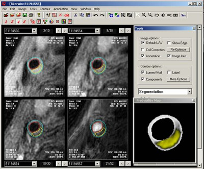
Plaque Segmentation with MEPPS
CASCADE features a patent-pending, histologically validated algorithm for multi-contrast segmentation of plaque components based on a combination of image intensities and plaque morphological features.
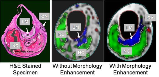
(see Liu et al. “Automated in vivo segmentation of carotid plaque MRI with morphology-enhanced probability maps.” Magn Reson Med 2006, 55:659-668.)
Vasa Vasorum Imaging
CASCADE features automated analysis of dynamic contrast-enhanced (DCE) MRI to assess plaque inflammatory markers. Vasa vasorum images highlight enhancement patterns that have been linked to neovascularization and macrophage infiltration of the plaque.
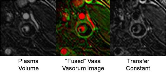
(see Kerwin et al. “MR imaging of adventitial vasa vasorum in carotid atherosclerosis.” Magn Reson Med 2008; 59:507-514.)
Validation
CASCADE has undergone rigorous validation for reproducibility, accuracy, and dependence on field strength.
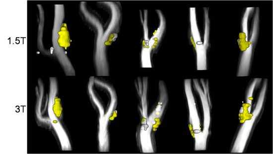
(see Kerwin et al. “Signal features of the atherosclerotic plaque at 3.0 Tesla versus 1.5 Tesla: Impact on automatic classification.” JMRI 2008; 28:987-995.)
Select VIL Publications Featuring Image Processing
- Kerwin et al. “Signal features of the atherosclerotic plaque at 3.0 Tesla versus 1.5 Tesla: Impact on automatic classification.” JMRI 2008; 28:987-995 .
- Kerwin et al. “MR imaging of adventitial vasa vasorum in carotid atherosclerosis.” Magn Reson Med 2008; 59:507-514.
- Kerwin W et al. “Magnetic Resonance Imaging of Carotid Atherosclerosis: Plaque Analysis” Top Magn Reson Imaging 2007; 18:271-278.
- Liu et al. “Automated in vivo segmentation of carotid plaque MRI with morphology-enhanced probability maps.” Magn Reson Med 2006, 55:659-668 .
- Underhill et al. “Automated measurement of mean wall thickness in the common carotid artery by MRI: a comparison to intima-media thickness by B-mode ultrasound.” JMRI. 2006; 24:379-387.
- Han et al. “Detecting objects in image sequences using rule-based control in an active contour model.” IEEE Trans Biomed Eng. 2003; 50:705-710.
- Kerwin et al. “Noise and motion correction in dynamic contrast-enhanced MRI for analysis of atherosclerotic lesions.” Magn Reson Med. 2002; 47:1211-1217.
- Han et al. A multi-scale method for automatic correction of intensity non-uniformity in MR images. JMRI. 2001; 13:428-436.
Acknowledgement
Support for this work was provided in part through an NIH grant (R44 HL070576) in partnership with VPDiagnostics, Inc.