


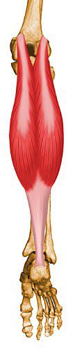
Origin: Medial head from posterior nonarticular surface of medial femoral condyle; Lateral head from lateral surface of femoral lateral condyle
Insertion: The two heads unite into a broad aponeurosis which eventually unites with the deep tendon of the soleus to form the Achilles tendon, inserting on the middle 1/3 of the posterior calcaneal surface
Action: Powerful plantar flexor of ankle
Innervation: Tibial nerve (S1, S2)
Arterial Supply: Each head supplied by a sural branch of the popliteal artery
The medical illustrations contained in this online atlas are copyrighted © 1997 by the University of Washington. They may not be utilized, reproduced, stored, or transmitted in any form or by any means, electronic or mechanical, or by any information storage or retrieval system, without permission in writing from the University of Washington.
Lower Extremity
- Adductor Brevis
- Adductor Longus
- Adductor Magnus
- Biceps Femoris – Long Head
- Biceps Femoris – Short Head
- Extensor Digitorum Longus
- Extensor Hallucis Longus
- Flexor Digitorum Longus
- Flexor Hallucis Longus
- Gastrocnemius
- Gluteus Maximus
- Gluteus Medius
- Gluteus Minimus
- Gracilis
- Iliacus
- Inferior Gemellus
- Obturator Externus
- Obturator Internus
- Pectineus
- Peroneus Brevis
- Peroneus Longus
- Peroneus Tertius
- Piriformis
- Plantaris
- Popliteus
- Psoas
- Quadratus Femoris
- Rectus Femoris
- Sartorius
- Semimembranosus
- Semitendinosus
- Soleus
- Superior Gemellus
- Tensor Fascia Lata
- Tibialis Anterior
- Tibialis Posterior
- Vastus Intermedius
- Vastus Lateralis
- Vastus Medialis