


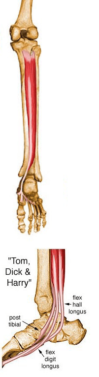
Origin: Posterior aspect of interosseous membrane, superior 2/3 of medial posterior surface of fibula, superior aspect of posterior surface of tibia, and from intermuscular septum between muscles of posterior compartment and deep transverse septum
Insertion: Splits into two slips after passing inferior to plantar calcaneonavicular ligament; superficial slip inserts on the tuberosity of the navicular bone and sometimes medial cuneiform; deeper slip divides again into slips inserting on plantar surfaces of metatarsals 2 - 4 and second cuneiform
Action: Principal invertor of foot; also adducts foot, plantar flexes ankle, and helps to supinate the foot
Innervation: Tibial nerve (L4, L5)
Arterial Supply: Muscular branches of sural, peroneal and posterior tibial arteries
The medical illustrations contained in this online atlas are copyrighted © 1997 by the University of Washington. They may not be utilized, reproduced, stored, or transmitted in any form or by any means, electronic or mechanical, or by any information storage or retrieval system, without permission in writing from the University of Washington.
Lower Extremity
- Adductor Brevis
- Adductor Longus
- Adductor Magnus
- Biceps Femoris – Long Head
- Biceps Femoris – Short Head
- Extensor Digitorum Longus
- Extensor Hallucis Longus
- Flexor Digitorum Longus
- Flexor Hallucis Longus
- Gastrocnemius
- Gluteus Maximus
- Gluteus Medius
- Gluteus Minimus
- Gracilis
- Iliacus
- Inferior Gemellus
- Obturator Externus
- Obturator Internus
- Pectineus
- Peroneus Brevis
- Peroneus Longus
- Peroneus Tertius
- Piriformis
- Plantaris
- Popliteus
- Psoas
- Quadratus Femoris
- Rectus Femoris
- Sartorius
- Semimembranosus
- Semitendinosus
- Soleus
- Superior Gemellus
- Tensor Fascia Lata
- Tibialis Anterior
- Tibialis Posterior
- Vastus Intermedius
- Vastus Lateralis
- Vastus Medialis