


This is unpublished
Findings:
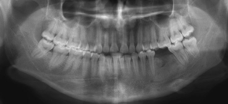
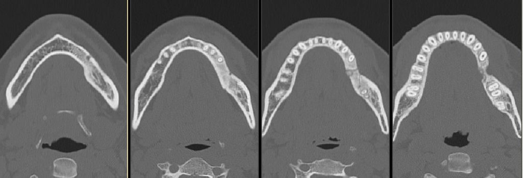
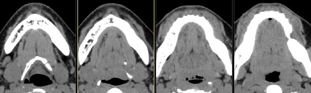
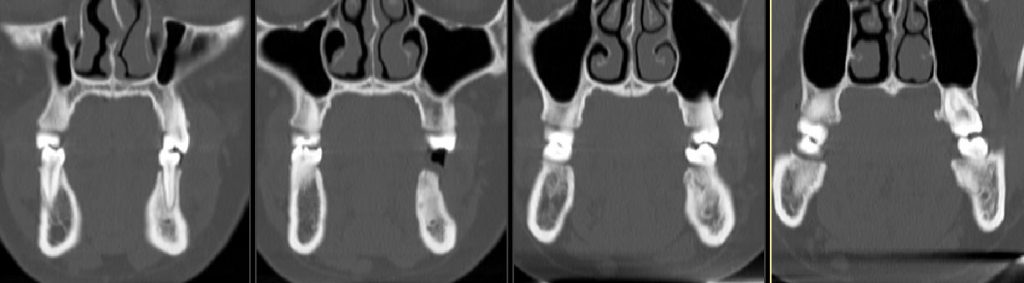
Diffuse sclerotic lesion of the body of left mandible with 5 mm nidus surrounded by lucent halo
DDX:
osteoid osteoma, osteoblastoma
Diagnosis:
Osteoblastoma of mandible
Discussion:
Osteoblastoma is a benign bone tumor accounting for 1% of all bone tumors; it commonly involves the spine and the sacrum of young individuals, with less than 5% being localized to the posterior mandible. Radiologically, they are usually poorly defined, radiolucent/radiopaque lesions containing calcifications and not showing sclerotic borders or periosteal reactions.
Submitted by Hari Challa, MD, UW Neuroradiology Fellow