


Menu
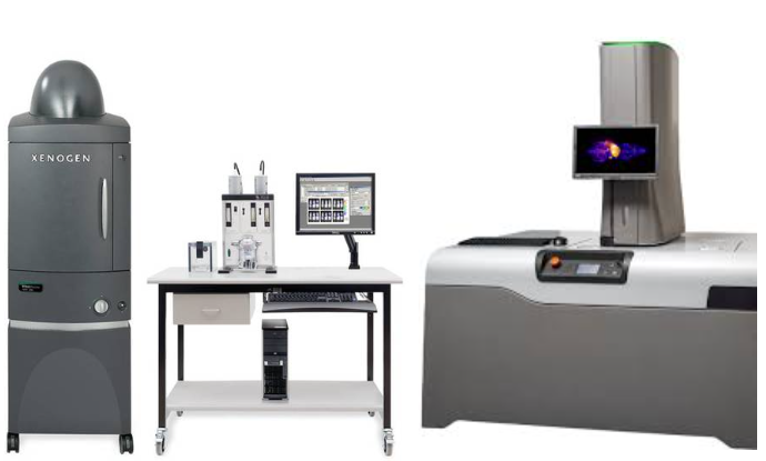
The Radiology Optical Imaging Core (ROIC) is equipped with the IVIS Spectrum In Vivo Imaging System and Xerra CFT System that is available to all UW labs as well as the surrounding research community. The IVIS is capable of performing optical in vivo imaging of bioluminescence and fluorescence in small animals, and the Xerra is useful for acquiring 3D fluorescent and anatomical data in small animals and tissues. Data acquisition for the IVIS is done using the Living Image software. The ROIC is located in the barriers at SLU E088B and ARCF B138.
Resources
Translational Bioimaging Core and Cancer Consortium
If you are interested in using the IVIS/Xerra, please contact Neal Paragas (paragas@uw.edu)

Neal Paragas, Core Director
Research Associate Professor
UW Radiology